Before diving in it would be useful to define what symptomatic and asymptomatic mean. In the last article on vaccine side effects, a specific set of symptoms including fever, cough, runny nose, malaise, achiness, mucus production (nasal and bronchial/lung), nausea, vomiting and diarrhoea were identified as being specifically related to activation and cascade of the immune system. These, and some other symptoms tell us, because of the mechanisms required to produce them, that the immune system is working well. ‘Side effects’ in this specific context represent immunity in action.
There can be a fine line, however, between desirable symptoms of detoxification and undesirable symptoms, indicating the body is overwhelmed with toxicity. and perhaps unable to trigger an immune cascade. These severe or fatal symptoms will be covered in a separate article.
But what is ASYMPTOMATIC? Arsine gas inhalation and radiation are both quite similar to carbon monoxide inhalation. We cannot see, taste, smell or sense it. Likewise, arsine and radiation are non irritant to mucus membranes, of course unless the dose is extremely high and acute (delivered at one time). The lack of irritation, and the immune system’s difficulty in detecting these invaders, means immune cascades are not triggered in the normal way. Arsine and the damaging effects of radiation build up in the tissues. Asymptomatic is represented by arsenic species embedding in proteins, or hitching a ride on an extracellular vesicle (EVs, exosomes etc) to other cells, or the silent breakage of double strands of DNA in the nuclei of cells.
It is only when cells start to die in large enough numbers that the immune system may realise something is wrong, and send in the cavalry. The signs of toxicity till that point may be non specific, a slight cough, dizziness, breathlessness or a nagging headache… The unproductive, dry nature of the cough indicating again that the immune system is not triggered, but that mucus membrane cells are being damaged. Breathlessness also exhibits with a silent collection of fluid in the lungs and other tissues, as endothelial cells are damaged and release a flood of interleukins and other cytokines. We may not be aware our lungs are full of this fluid until we have difficulty catching our breath, or until blood oxygen saturation is so low urgent medical intervention is needed. Asymptomatic testing would be a useful in identifying excessive levels of toxicity and/or radiation. This would lead to exposure reduction through quarantine, and turnover of contaminated cells during this period, all allowing tissue levels to drop.
In this article when we refer to arsenic, we are not forgetting the damage other toxic elements such as Cadmium, Mercury and Lead can cause to cells.
Problem number 1 – Recognising the Enemy
This is where the whole concept of ‘Coronavirus’, or even viruses in general, gets very ropey. We are told that the SARS CoV 2 virus is 30,000 amino acids long with spike proteins each 1,273 amino acids long. The whole vesicle measures approximately 120 nanometres across. A large virus by all accounts. Titin, the longest protein in the human body is up to 35,000 amino acids long. How on earth is it possible, with a virus that size, that the human body did not detect it as a foreign protein at some point between entry in to the respiratory system and the widespread hijacking of cellular machinery and send immune cells to kill it? Shhhh, what’s that? Yes, we will come to capsids.
Taking this idea further, in the theory of virology, initial exposure to a pathogen is meant to prime the body to recognise the pathogen a second time. Immune system priming is covered in this article here. We have established that a significant number of people, in the UK at least, experienced what we now know to be Covid-19 towards the tail end of 2019. Therefore, if priming the immune system theory is applied, all of these people should have been able to mount a suitable defence on future exposures. This also goes for those who were ‘infected’ in the first wave. Immunity to the pathogen should have been widespread by that point and herd immunity almost achieved, as many believed Boris was gunning for. Yet for some reason scientists felt that a vaccine was still required. A small note on variants, please refer to the section on viral sequencing below.
The vaccines were to deliver, supposedly, the spike protein part of the larger virus for the body to replicate so that it could mount a defence against it. But if our bodies struggled to mount a defence against a Titan (Titin) sized ball of protein, what hope did it have of mounting a defence against a spike protein 23 times smaller?
Problem number 2 – Codons and Amino Acids
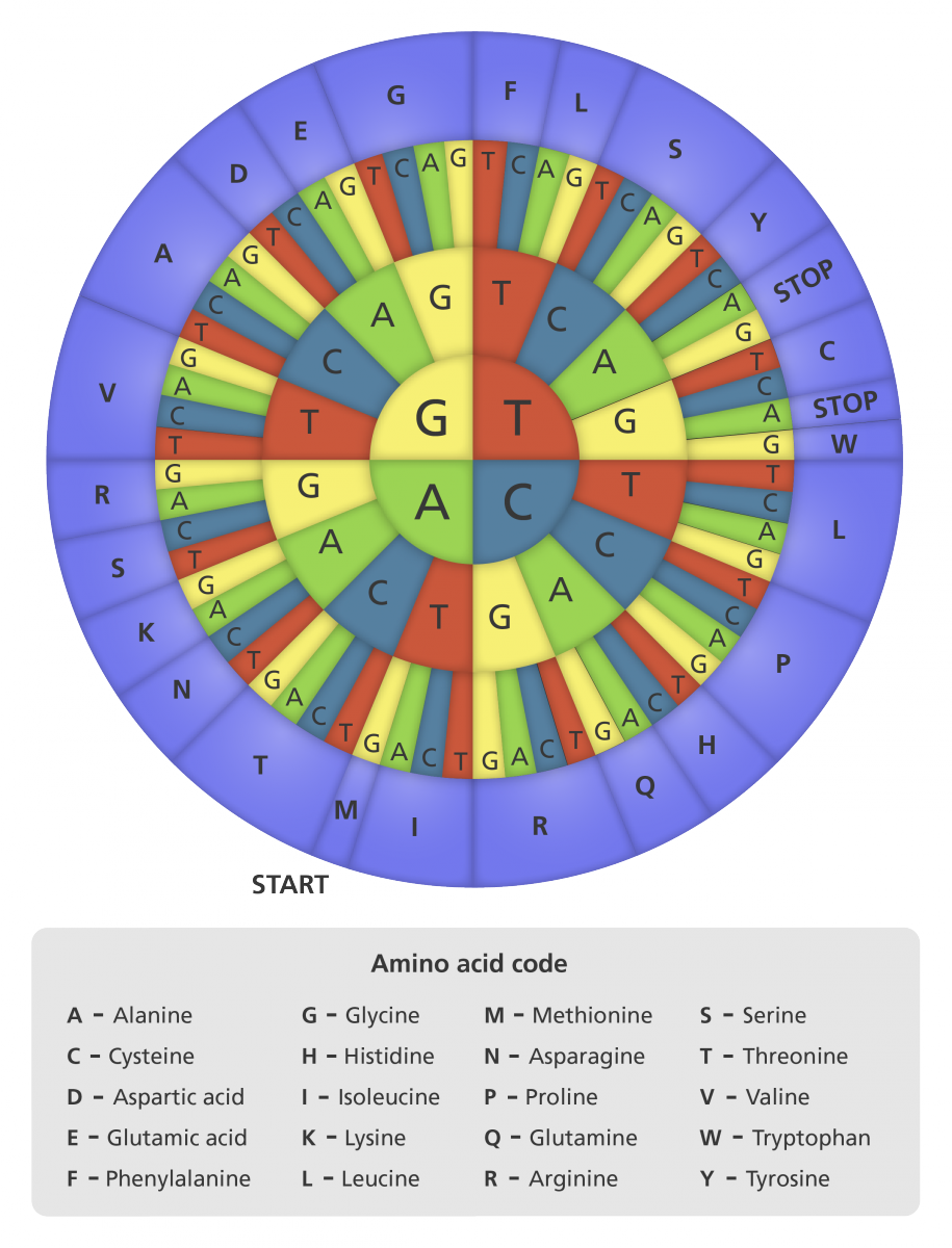
All organic matter is made up of 20 amino acids and combinations of codons. There are only 64 codons and arranged in triplets make up the 20 amino acids. One amino acid can have a few combinations of codons as seen below in this chart. This means 64 codons make up all the protein chains in all living organisms.
When we tried to decode the SARS CoV 2 spike protein from end-to-end, using only human amino acid peptide chains, it was a great surprise to find that we could. We found multiple possible human proteins for peptides between 4-7 amino acids long. We invite others to repeat the challenge. Let us know what you find.
We also found, in this three day exercise, that some human protein/peptide results had very few species crossmatches. In some cases the only match was SARS CoV 2, sometimes SARS CoV and perhaps a yeast or mouse protein. The database only returned a human protein to match a peptide query together with SARS CoV 2 protein and other organisms – it was never on its own.
In this short article on how to find a ‘virus’, researchers from the Perelman School of Medicine in Philadelphia actually hunted through tissues in the human body to find free floating viral particles. From these they ‘identified short sequences that were similar to a type of virus found in domesticated pig stool’ (that’s pig poop). But what did they mean by ‘short sequences’? The same short sequences of 4-7 amino acids that are present both in human and the SARS CoV 2 proteins? Together with our findings on lack of protein and peptide duplication over approximately 1390 protein options it all seems very strange!
Consulting a molecular biologist friend, they also agreed the lack of repetition and the SARS CoV 2 affinity with so many sections of different human proteins was very strange. To which it seemed sensible to propose that perhaps ‘virus’ as we know it may not actually be a long foreign strand of DNA or RNA, that actually it may just all be jumbled up snippets of human DNA and RNAs. The consensus was, that although it had not been thought of like that before, it was entirely possible.
Problem number 3 – Viral Sequencing
In the past it was rare for an entire viral genome to be sequenced. Viruses were identified by certain characteristics, rather than the exact imprint of the nucleotide chain. Indeed in the above study by Perelman School of Medicine, the researchers knew their findings were viruses apparently due to their similarities to previously discovered viral molecules.
In ‘A new methodology for sequencing viruses’ (Phys.org, 13.09.2019) isolation of viral genomes seems fraught with problems. The samples are often far too contaminated with human genetic material. Researchers are often unable to ‘find’ the virus unless they know something about characteristics it has already, in order to extract what they believe to be the virus from the other human material. This implies there is a level of prediction involved in identification of ‘viruses’.
What these inherent similarities also imply where variants are concerned, is that certain characteristics will remain the same, and identification and treatment should be able to overcome small variations.
In summary we have a human and supposedly viral soup, all jumbled up, in which scientists try to find a small sequence or two that they recognise as a previously identified viral snippet. In this experiment detailed above they, then design little traps, very possibly using these snippets, to supposedly attract the complete ‘viral genome’ away from the human material to ‘isolate’ it. Are you thinking what we are thinking?
When we were shown the supposed ‘viral’ affiliation of Covid-19 to bats, HIV and other genomic sequences, the alarm bells should have started to ring. 64 Codons and 20 amino acids is not a lot of material to play with. Probability says there is going to be significant repetition across species of proteins and peptides. There has to be!
Let’s imagine scientists managed to extract 40 viral particles at exactly the same time, from the same human and sequence them simultaneously. Would each virus particle be the same end to end? It is possible that each of these viral particles may have some common characteristics, but the remaining material might be quite different. But it would only be evident if they sequenced all 40 at the same time.
Problem number 4 – Mismatched bases
Testing for viruses uses a similar principle to identification of ‘viruses’. Scientists provide templates for mass testing which feature the nucleotide sections that they witnessed the most often in that particular ‘virus’, not the entire virus. When samples of material from human test subjects are passed through multiple stages of PCR amplification, sometimes the primer and the template DNA or RNA mismatch. This may or may not cause problems in amplification. The outcome of a small mismatch could be that something identified as something foreign, might actually be human.
There are also problems in choosing a string of bases that is absolutely conserved, or completely stable and unchangeable. In this article by Bru et al. (2008) discussing the ‘…Detrimental Effect of a Single Primer-Template Mismatch by Real-Time PCR…’ they find in the observed bacterial gene (16S rRNA gene) ‘the longest string of totally conserved bases is between positions 788 and 798, and most of the absolutely conserved regions are found in strings of less than 4 bases’, In other words, the only stable parts of the gene being tested are really very short indeed.
In summary a single mismatched base can totally throw off the accuracy of a PCR amplification, depending on where it is in the chain.
Unless of course that is the PCR isn’t testing for a highly mutable amino acid sequence, as the number of Covid-19 variants demonstrates, but instead for a fixed marker such as Gamma-H2AX, indicating a DNA double strand break as discussed in this article.
This, together with the structural and content similarities between viruses and the broad range of human extracellular vesicles, lends weight to the possibility they are all slight variations of the same thing, vesicles budded from the cell wall via an endosome. Albeit the ‘virus’ extracellular vesicle may well be a vector for the spread of arsenic! It is also possible that the contents of said ‘virus’ do not look like human DNA or RNAs, as they have been sheared by radiation or arsenic, and potentially reassembled in to proteins that bear little resemblance to human proteins.
Problem 4 – THE CAPSID
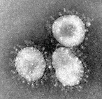
Where is the capsid in all this soup? Here is a picture of a coronavirus from an electron microscope. Surely it would be easy to identify a ‘virus’ from that alone, unless of course there is human material also in the ‘virus’. Can you see a polygonal structure in this image?

As researchers struggle to find the ‘viral genome’ in amongst a soup of human material, the viral capsid becomes even more of a question mark. A capsid is a protein sheath forming either a rod helical (spring) shape around the viral genome or a ball-shaped symetrical polygon around the viral genome. In the electron microscopy image above of Coronavirus, the polygon capsid is not visible, and we have been unable to find a real image that shows this structure. This image from Getty apparently shows the measles virus in great detail, but again the symetrical polygonal structure seems to be missing as the lipid shell is degrading.
What looks like a polygonal capsid, might be the inner section of the lipid bilayer present in extracellular vesicles, at a certain point in time. If the vesicle has been contaminated with arsenic species, or other toxic material, it is possible that the inner part of the bilayer has crenated. This is where the walls of a cell become leaky, due to toxin activity. The walls begin to pucker inwards as seen in the photo here. As the toxin is on the inside of the vesicle (virus), the crenation may occur from inside-out, making the inner lipid layer look polygonal, before it reaches the outer lipid layer.
Then of course we still need to consider the difficulties researchers have in separating viral material from human, when the virus is as identifiable as the photo featured above. What is really inside that vesicle?
mRNA Quality Control
There are various checks and balances during the copying from DNA to create pre-mRNA, and copying of mRNA by ribosomes to produce final proteins. Sinchen (Susan) Shao, Harvard Medical School, gave a talk ‘Mechanisms of ribosome-associated quality control’ two months ago to the Broad Institute about nascent (new) protein synthesis. Her and her team’s focus was on how aberrant (faulty or foreign) mRNA is identified.
Ribosomes communicate with each other about events and features that compromise quality control. The mRNA must have start and stop codons, not include premature stop codons, and sometimes there will be translational stalling due to stem loops. These three represent the main pathways of mRNA quality control when it reaches the ribosome for translation, and can trigger decay if faulty mRNA is detected. Any aberrant mRNA trying to make it to the end of translation without decay, must rely on all of these quality control mechanisms being broken, without the ribosome function being altered, or somehow the ‘virus’ being able to mimick human mRNA to the extent that all of these identifying markers are in the correct places. A very tall order.
In virology we are told that viruses hijacked cellular machinery, preventing human mRNA from being transcribed. There are a few considerations here. The initial steps in the creation of mRNA may be disabled, not by the virus, but by arsenic contamination. To see if that is likely we looked at some of the proteins involved.
RNA polymerase enzyme binds to the promoter section of DNA. Professor Dave explains the transcription and translation of DNA to mRNA perfectly in his video here. The binding attaches ribonucleoside triphosphate to the polymerase. As the mRNA has already moved out of the nucleus in to the cytosol to bind with the ribosome, the triphosphate involved in this process is potentially vunerable to arsenate substitution. Zinc is also involved in stabilisation of RNA polymerase (I). DksA, a polymerase accessory protein for regulation and sensing oxidative stress, has a 4-cysteine zinc-finger motif. Zinc is usually bound to cysteine amino acids, and cysteines with their sulphur atoms are a target for arsenic species. Attachment or displacement by arsenic species could inactivate all of these processes, stalling the translation before it has even got off the starting block.
Cleavage factors which process 3’ (three prime) end machinery of the pre-mRNA are all phosphoproteins. Phosphotyrosein has a phosphate type group (orthophosphate) which may be substituted by arsenate. Cleavage and polyadenylation specific factors and cleavage stimulation factor complexes contain phospherine, which also have phosphate groups attached.
There are many factors and enzymes involved in the transcription of pre-mRNA from DNA. All those containing cysteine or phosphate groups are targets for arsenic species. If the vesicle carrying arsenic contaminated proteins arrives in the cell, these could easily contaminate parts of mRNA machinery. In effect it really does look like it is turning transcription processes off in the nucleus!
A new theory
For this next part if you are a microbiologist or virologist, please bear with us, this is an entirely new thought process and of course it will lack in concrete evidence, but perhaps the mechanism might seem feasible enough for one of you to investigate.
Let’s imagine that a cell has lost the ability to produce mRNA from it’s own DNA because of this arsenic contamination. Also let’s imagine it and it’s neighbouring cells are all panicking. They are struggling with contamination, their inability to make mRNA to maintain cell integrity and from damage to other cellular components. In their panic they throw out a number of extracellular vesicles packed with RNAs, DNA and other proteins (this is what virologists might call a cell making/shedding more virus particles). These protein parcels may contain proteins that warn healthy cells about the incoming contamination chaos, or tools to fix the issue or help the cell defend against the problem. They cannot detect, as our immune systems cannot, the presence of arsenic species attached to their cargo.
More chaos ensues as the arsenic is unloaded in the target cell. See this article again for how many of the surface proteins of EVs include disulphide bridges, which are like candy to arsenic. Very quickly machinery in target cells will become faulty and a similar breakdown will occur. The target cell will also send out warning and fixer proteins to its neighbouring cells, spreading the contamination further.
This paper on ‘Cell to Cell Signalling via Exosomes Through esRNA’ by Lotvall & Valadi (2007) shows clearly that extracellular vesicles specifically identified as exosomes can deliver fully functional RNA and also ready-to-copy mRNA to target cells. In the case of our arsenic laden EVs, there may well be some non-contaminated mRNA which is viable, and possibly also functional RNA. Proteins affected by arsenic may not be translated. They may have been broken or even attached via cysteine molecules to the cysteine molecules in other proteins in the vesicle. They may have lost the correct start and stop sequences, or the correct Exon Junction Complex spacing. These junctions, spaced out along the mRNA chain, are what indicate to the ribosome and other copying machinery that the mRNA protein is human, and has been represented accurately.
So despite the cell’s inability to make mRNA directly from the nucleus, it may still be able to translate some of the mRNA proteins received in the extracellular vesicles. It may appear to observing scientists (who are not in the habit of testing for arsenic contamination), however, that somehow these ‘viruses’ or EVs have taken over the cell’s remaining active ribosomes AND have somehow ground DNA transcription to a halt. This supposed ‘hijacking’ and switching off of pre-mRNA machinery is a characteristic virologists believe is intrinsic to the behaviour of viruses. As arsenic is so incredibly difficult to detect, as it merges with proteins, it is quite understandable that it may have been missed.
Using an analogy, the cell of origin has DNA and proteins, a bit like a long newspaper article on Megan and Harry. The article can be read and we understand its meaning. When arsenic (and radiation) gets involved, some of those proteins become broken, adhered to other proteins they should not be and everything ends up a whole jumbled mess. It would be like cutting up the long article about Megan and Harry making fun of the Royal Family in to short 4-7 word phrases, putting them in a bowl and picking out 30 snippets and trying to piece them together in to something intelligible. The final message says something along the lines of Megan and Harry are really sorry for the distress they have caused to the family and Royal followers around the world, they will do a suitable amount of time out from the headlines as punishment. The real issue, like in the rearrangement of these phrases, may have been completely lost in translation.
Human mRNA?
So by this point we, and possibly you, are pretty sure that what is called a ‘virus’ probably isn’t, and that toxin contamination of cells via intercellular messaging is a large part of the picture. So if the ‘virus’ isn’t really a virus, and the proteins in and on extracellular vesicles are just human proteins, possibly contaminated with arsenic, what is the mRNA in the vaccines?
mRNA is not something that you can just stick in to a human cell, for the ribosome to produce proteins from. It must have all the attributes of human mRNA as listed above. If it is delivered complete with lipid outer layer to the cytoplasm ready for copying, it must still have start and stop codons, and no premature stop codons or stem-loops. For these reasons we believe it is most likely, if mRNA is being delivered and not a complete oven-ready protein, that the mRNA chosen for the vaccine is of human origin.
The signs of detoxification in many vaccine recipients lead to two types of protein candidate. One being a protein, possibly an enzyme, that triggers immune pathways in a way that the Covid-19 ‘virus’ didn’t, towards a generalised autophagy. The second would be a protein that physically cleans up any arsenic from the cells, and by their increased presence, actively promotes an immune cascade. It might well be that there is more than one type of mRNA in each lipid capsule, one to capture the enemy, and another to see it to the door. More to come…
Thanks Seb, we are getting there!


